
What Is An Ultrasound Transducer ?
A ultrasound transducer is a device that can converts one from of energy in to other form. Transducers are used to convert an electric signal into ultrasonic energy which can be transmitted into tissues and to convert ultrasonic energy reflected back from the tissue into an electric signal. It works on the principle of piezoelectric effect. It is a hand-held device that is placed directly on the skin or within a body cavity, and it emits sound waves that penetrate the body and bounce back to the transducer.
The transducer then converts the echoes into electrical signals, which are processed by a computer to produce images of internal organs, tissues, and blood vessels. Ultrasound imaging is non-invasive and does not use ionizing radiation, making it a safe and widely used diagnostic tool in medicine.
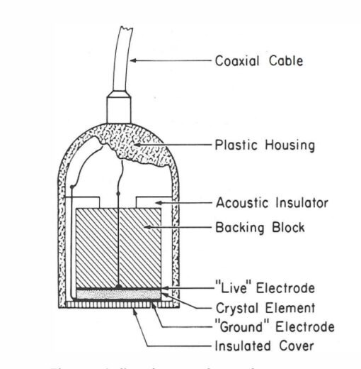
Structure Of Ultrasound Transducer.
In 1880 Pierre and Jacques Curie first observed the piezoelectric property in quartz, Rochelle salt and tourmaline. The transducer works on piezoelectric effect principle. The transducer elements are 6 – 9 mm in diameter and 0.2 to 2 mm thick. The transducer elements are generally curved for better focusing by may be flat in shape.
Major components of transducer are :-
A) the piezoelectric material.
B) matching layer.
C)Backing Material and Damping.
D) acoustic absorber.
E) Housing ( plastic or metal ).
F) Cable and connector.
A) Piezoelectric material:- The active element of the transducer is made up of one or more piezoelectric crystals, which are usually made of a ceramic material such as lead zirconate titanate (PZT). It converts electrical energy into mechanical (sound) energy by physical deformation of the crystal structure. The crystals are shaped and cut to a specific size and thickness depending on the desired frequency of the sound waves they will produce.
B) Matching layer:- The matching layer provides the interface between the transducer element and the tissue and minimizes the acoustic impedance differences between the transducer and the patient. The thickness is equal to one – fourth the wavelength, which is known as quarter wave matching.
C) Backing Material and Damping:- The backing material, as the name implies, sits on the back of the transducer, behind the elements . The purpose of the backing material, also referred to as the damping material, is to provide damping of the piezoelectric element. The damping material serves to shorten the length of the pulse by decreasing the number of cycles in the pulse. This material is composed of an epoxy resin loaded with tungsten.
D) Acoustic absorber:- A curved piece of material, such as plastic or rubber, is placed in front of the piezoelectric crystals to focus the sound waves into a narrow beam. The shape and thickness of the lens can vary depending on the desired beam width and depth of penetration.
E) Housing ( plastic or metal ):- The components of the transducer are housed within a protective casing made of plastic or metal. The housing may have a tapered shape to allow for easy maneuverability during imaging.
F) Cable and connector:- The piezoelectric crystals are connected to the ultrasound machine via a cable, which is typically shielded to prevent interference from external sources. The connector is usually a threaded or bayonet-style connection that ensures a secure connection between the transducer and the ultrasound machine.
Types Of Ultrasound Transducer.
Transducers that give real-time imaging of the anatomy and physiology of body organs are called real-time transducers. Real-time ultrasonic images use two basic techniques which are:
A. Mechanical Transducers.
B. Electronic Transducers.
A. Mechanical Transducers:- These transducers typically had one or more piezoelectric elements connected to a motor, or a fixed element with a mirror connected to a motor. The motor, or a mirror, steered the element to produce the scan lines that made up the image. This produced a sector image pattern. The piezoelectric element and motor were inside of a protective housing. Oil was used as a coupling medium to prevent air from forming within the housing. Air within the housing would hamper the transmission of the sound. These transducers were fixed frequency and fixed focus. That is, in order to change the frequency or the location of the focal zone, one had to change the entire scan head. Focusing of the beam was achieved by either the shape of the element or the use of a lens.
The major advantages of the mechanical transducer were that they were inexpensive and typically had a small footprint. These transducers were fragile and their mechanical elements were easily broken.
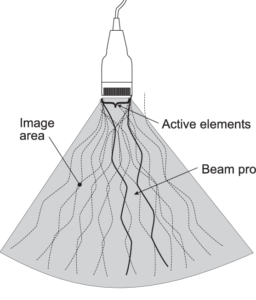
B. Electronic Transducers:- Electronic scanning is performed with transducers that have multiple active elements. This is referred to as an array. An array is formed by taking a single slab of PZT and slicing it down into multiple sub elements. Each sub element is connected to a wire, so it may fire independently. The system can selectively excite the elements as needed to shape and steer the beam. With most array transducers, no motors are needed for beam steering. Arrays may be either sequenced or phased and can produce various image shapes.
Following types of electronic transducer.
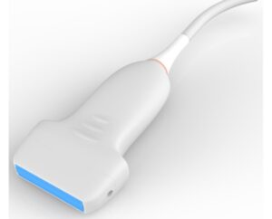
- Linear Array:- This type consists of a number of small rectangular transducer elements arranged in a line. The linear probe is a high-frequency probe. The piezoelectric crystal arrangement of the linear probe is linear. The beam shape of the linear probe is rectangular, and it has a good near-field resolution. The 2D imaging linear transducer has a wide footprint and it has a frequency ranging from 2.5 MHz to 12 MHz. It is the best probe to view the superficial structures. It provides information about poor depth organs, and it has low penetration power.
Uses of Linear Probe:
It is used for viewing:
— Vascular examination
— Blood Vessel visualization
— Musculoskeletal – tendons, bones, muscles
— Eyes
— Breast
— Thyroid Gland
— Tendons
— Testicular
— Appendicitis
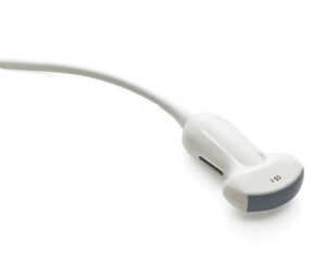
2. Curved Array :- The curved sequenced array transducer, also referred to as a convex, curvilinear, or curved sequential array, is based on the same technology as that of the linear sequenced array but with a curved face . As with the linear sequenced array, the elements are fired in groups.
The shape of its beam is convex and becomes wide at the footprints. These probes have frequencies ranging between 2.5 to 7.5 MHz. the sub-type of the curvilinear probe is called the micro-convex which has a smaller footprint and due to this reason, it is used in neonatal and pediatrics.
3. Phased Arrays:- The phased array is more commonly known as a sector or vector transducer. The sector/vector transducer typically has a small footprint, also referred to as the “face” of the transducer, and it may be used for cardiac imaging, neonatal brain imaging, with some endocavitary transducers, and any other application where a sector or vector image shape is desired . In order to create a sector image, electronic steering is needed for every scan line. Unlike the curved and linear sequenced arrays, where the shape of the transducer dictates the shape of the image, in the phased array the shape of the face of the transducer does not resemble the shape of the image.
This transducer can make either the sector, which yields a true “pie-shaped” image, or the vector image shape, which yields the flat-topped, trapezoidal image shape . With the sector phased array transducer, all of the scan lines originate from a common point of origin. The phased array transducer uses phasing to steer and focus the beam. Phasing is changing the timing of the shocking of the elements in order to shape and steer the beam (Figure 2-20). All arrays are phased focused. That is, all of the array transducers mentioned in this section use phasing to control the focusing in the scan plane. Phasing provides the sonographer the ability to control the depth of the focal zone in the scan plane. The order in which the elements are shocked determines beam steering and focusing.
FOR MORE ULTRASOUND CLICK HERE
BOOK LINK :- Understanding Ultrasound Physics