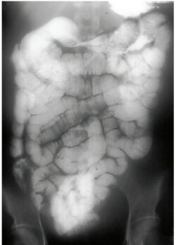
What Is Small Bowel Enema ( Enteroclysis)?
The small bowel enema is the radiological examination to help the study of the small bowel from jejunum to ileocecal junction by intubation of contrast media through the Bilbao dotter tube in jejunum. It is also called Enteroclysis.
Indication of Small Bowel Enema ( Enteroclysis)
— Abdominal pain
— abdominal mass
— Intestinal tuberculosis
— Intestinal obstruction
— Intestinal polyps
— Inflammation
— Diarrhea
— Intestinal bleeding
— Carcinoma
— Weight loss
— Intestinal stricture
— Intestinal Perforation
— Diverticulum
— Malabsorption
Contraindications 0f Small Bowel Enema ( Enteroclysis)
— Suspected perforation
— paralytic ileus
— Completely obstruction
— Recurrent abdominal surgery
Contrast media.
— 20% W / V suspension of barium sulphate solution.
— 200-250 % W /V suspension barium sulphate solution.
Equipment.
— Fluoroscopy unit.
— Spot film device.
— Bilbao dotter tube.
— Local anesthesia.
— Carboxylic cellulose.
Patient preparation.
— The patient must be NPO 5 – 6 hrs. before the examination.
— The patient must be give the instructions to take low diet for one to two day before the examination.
— Laxative may be given to patient to remove the all feces material from the abdomen.
— The patient must be stop smoking or tobacco for prevent the proper coating of barium sulphate on the mucosal.
Procedure / Technique.
When the patient come in radiology department.
— To ask the patient to remove all the clothes and wear the hospital gown.
— To explain the whole Procedure before the patient.
— Take the consent from to patient or patient attender.
— The patient sits on the edge of Fluoroscopy table with head is extended.
— The local anesthesia spray apply in patient pharynx to sedation and the local anesthesia jelly apply on the Bilbao dotter tube.
— Then the tube is inserted through the mouth or nose.
— The patient is asked to swallow as the tube passed through the gastric antrum.
— Then the patient in supine position and the tube passed into the duodenum .
— When the Bilbao dotter tube are in jejunum then the guide wire is removed.
SINGLE CONTRAS METHOD.
— The low density barium sulphate solution 29% W / V suspension is inserted continue as help of Bilbao dotter tube under the observation of Fluoroscopy.
— When the barium sulfate solution are coating the layers of loops then the spot film taken.
DOUBLE CONTRAST METHOD.
— The high density of barium sulphate solution 250%W/ V used in double contrast method.
— The barium sulphate solution inserted continuously in jejunum through the Bilbao dotter tube.
— After reaching the contrast in jejunum the methylcellulose is insurted in jejunum.
— Then the spot film are taken.
Filming
AP view:- Full abdomen.
— PA prone view:- For bowel pressure
— Oblique:- For separate bowel loops
— Erect:- For evaluation of fluid leveling diverticulum .
Aftercare.
— Ask the patient to take a maximum fluid.
— To inform the patient about feces will be white for some days.
— Gave the Dulcolax to remove the barium sulfate solution through the feces.
Complications.
— Leakage barium sulphate solution for perforation.
— Aspiration.
— It may caused appendicitis.
— Acute gastric dilatation.
— Barium impaction.
ALSO READ CLICK HERE
BOOK LINK : RADIOLOGICAL PROCEDURES, 2E

