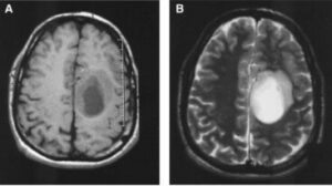
What Is Oligodendroglioma?
Oligodendroglioma is a type of brain tumor that arises from oligodendrocytes, cells that produce the protective myelin sheath around nerve cells in the brain. It is a slow-growing, low-grade tumor that occurs most commonly in the cerebrum. Symptoms may include seizures, headaches, vision changes, and changes in behavior or personality. Treatment typically includes surgery, radiation therapy, and chemotherapy.
Oligodendrogliomas are relatively rare, accounting for about 2-5% of all primary brain tumors. They typically occur in adults, with the average age of diagnosis being around 40 years old. Oligodendrogliomas are generally slow-growing tumors that tend to remain confined to the brain and rarely spread to other parts of the body.


What Are The Types Of Oligodendrogliomas?
There are three main types of oligodendrogliomas:-
A. Grade II oligodendroglioma:- This is the most common and slow-growing type. It is considered a low-grade glioma. Tumor cells look somewhat abnormal under the microscope but not very aggressive. About 70-90% of patients survive 5 years with treatment.

B. Anaplastic oligodendroglioma (grade III):- This is a higher grade, more aggressive form of oligodendroglioma. The tumor cells look very abnormal and are actively dividing. Prognosis is worse, around 25-50% 5-year survival. This can develop from a grade II oligodendroglioma over time.

C. Oligoastrocytoma (grade II-III):- This is a mixed glioma that contains both oligodendroglial and astrocytic tumor cells. It has an intermediate aggressiveness between oligodendroglioma and astrocytoma. Grade II has 60-90% 5-year survival while grade III is around 35-55%.
What Are The Symptoms Of Oligodendrogliomas?
The symptoms of oligodendrogliomas can vary depending on the location of the tumor in the brain and the size of the tumor. Some of the common symptoms include:-
— Headaches.
— Seizures.
— Cognitive and memory problems.
— Weakness or numbness.
— Vision or speech problems.
— Behavioral changes.
— Nausea/Vomiting
— Memory loss
— Cognitive impairment
— Coma
— Difficulty speaking
— Muscle weakness
How Are Oligodendrogliomas Diagnosed?
The diagnosis of oligodendroglioma typically involves the following tests:-
A. Imaging scans:- The most common scans for diagnosing oligodendroglioma are MRI, CT scan and PET scan. These can detect the tumor location, size, growth pattern and extent of infiltration. MRI is the most sensitive for detecting these brain tumors.


B. Biopsy:- Surgical removal of part of the tumor tissue (biopsy) is usually needed for an accurate diagnosis. The tissue sample is analyzed under the microscope by a neuropathologist to determine the tumor grade, cell type, genetic mutations and other features.
C. Histopathology:- Examining the tumor cells and tissue under the microscope is critical for diagnosis. Oligodendrogliomas have a characteristic histologic appearance with isomorphic cells, perivascular processes, and reticulin network. Grade determines how abnormal the cells look.
D. Immunohistochemistry:- Special stains are used to determine if the tumor is primarily oligodendroglial, astrocytic or mixed. Antibodies against olig2, AGC and GFAP proteins help identify oligodendroglioma.
E. Genetic testing:- Analyzing the tumor DNA for genetic mutations like co-deletion of 1p and 19q chromosomes is very useful for diagnosis and prognosis of oligodendroglioma. These mutations are present in about 80% of grade II-III oligodendrogliomas.
F. Neuropsychological evaluation:- For some patients, meetings with neurologists, neuropsychologists, speech pathologists and occupational therapists may be needed. They evaluate cognitive, behavioral, language and motor skills which can be affected by an oligodendroglioma.
G. Lumbar puncture:- In rare cases, a needle is inserted into the lower spine to remove cerebrospinal fluid for analysis. This checks if any cancer cells have spread into the fluid from the tumor.
H. Other tests:- Blood work, EKG, bone marrow biopsy, etc. may be done depending on the tumor grade and patient’s symptoms/condition. But they are not routinely needed for diagnosis of oligodendroglioma.
What Are The Treatments For Oligodendrogliomas?
The treatment of oligodendrogliomas depends on several factors, such as the location and size of the tumor, the age and overall health of the patient, and the tumor’s genetic characteristics. The following are some of the common treatments for oligodendrogliomas:-
A. Surgery:- The goal of surgery is maximum safe removal of the tumor while preserving brain function. The tumor is resected as much as possible, while avoiding damaging important brain areas like centers for vision, speech, movement, senses, etc. Even partial removal can provide some relief from symptoms.
B. Radiation therapy:- External beam radiation therapy uses high-energy beams to kill any remaining tumor cells after surgery. This helps control regrowth of the tumor. Radiation can be recommended especially for higher grade oligodendrogliomas.
C. Chemotherapy:- Chemotherapeutic drugs are used to destroy tumor cells that may not be eliminated with surgery and radiation alone. Temozolomide is a common chemo drug for oligodendroglioma. Chemotherapy may provide additional benefit in terms of longer progression-free survival, especially for co-deleted 1p/19q types.
D. Targeted drug therapy:- Newer targeted drugs are being investigated which block specific genetic mutations driving oligodendroglioma growth. These include PI3K inhibitors, MEK inhibitors, mTOR inhibitors, etc. Some are showing promise in clinical trials.
E. Clinical trials:- Participating in clinical trials for new treatment approaches such as immunotherapy, nanotechnology, etc. can provide access to innovative therapies for patients with oligodendroglioma. Many trials are evaluating targeted drugs, vaccines, and other new options.
F. Supportive care:- Managing symptoms and side effects is an important part of oligodendroglioma treatment. This includes managing seizures, corticosteroids for swelling, pain relievers, nutrition support, etc.
FAQs.
Q. What is Oligodendroglioma?
A slow-growing primary brain tumor that develops from oligodendrocytes, which produce myelin insulation for nerve fibers.
Q. What are the grades?
Grade II is low-grade, Grade III is anaplastic (more aggressive). Most oligodendrogliomas are Grade II.
Q. Where does it occur?
Oligodendrogliomas typically arise in the frontal, parietal or temporal lobes. They are intra-axial tumors.
Q. Who gets it?
Mostly adults aged 30-50 years. No known risk factors but some genetics links.
Q. Is oligodendroglioma brain cancer?
Grade III oligodendrogliomas are malignant (cancerous).
Q. What is the survival rate for oligodendroglioma?
About 30 to 38% of people with this type of tumour will survive for 5 years or more after they are diagnosed.
Q. Can oligodendroglioma be cured?
Approximately one third of patients with oligodendroglioma appear to be cured with aggressive treatment.
Q. What is the longest survival of oligodendroglioma?
The longest survival reported was 35 years
Q. Is oligodendroglioma malignant or benign?
Oligodendrogliomas can be malignant (cancer) or benign (not cancer).
Q. Can a oligodendroglioma grow back?
Prognosis. Oligodendroglioma is currently an incurable disease and the chances that it will recur and eventually progress are very high
Q. Is oligodendroglioma genetic?
The etiology is not known but no hereditary forms of oligodendroglioma have been reported.
FOR MORE UPDATES:- CLICK HERE
BOOK LINK :- Brant and Helms’ Fundamentals of Diagnostic Radiology

