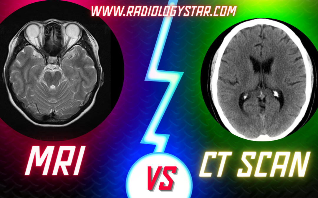
What Are MRI Vs CT Scan
IN MRI Vs CT Scan, Medical imaging plays a crucial role in diagnosing and monitoring various health conditions. Among the most common imaging techniques are Magnetic Resonance Imaging (MRI) and Computed Tomography (CT) scans. While both provide valuable insights into the human body, they are fundamentally different in terms of technology, applications, and radiation exposure. In this article, we will delve into the world of MRI and CT scans, exploring their principles, strengths, limitations, and the situations in which each is the preferred choice. By the end, you will have a clear understanding of when to use MRI vs. CT scan, ensuring the best care for your health.
Magnetic Resonance Imaging( MRI )
Magnetic Resonance Imaging (MRI) is a powerful medical imaging technique that utilizes strong magnetic fields and radio waves to create detailed images of the body’s internal structures. Unlike CT scans and X-rays, MRI does not use ionizing radiation, making it a safer option for repeated imaging and for certain patient populations, such as pregnant women and children.
MRI’s ability to produce high-resolution, cross-sectional images makes it an excellent choice for examining soft tissues. It excels in visualizing the brain, spinal cord, muscles, joints, and organs like the liver, kidneys, and heart. MRI provides exceptional contrast between different types of soft tissues, allowing for precise localization of abnormalities.
One key advantage of MRI is its capability to capture dynamic processes, such as blood flow and the movement of cerebrospinal fluid. Functional MRI (fMRI) can even map brain activity by monitoring changes in blood flow.
Principle:
— MRI relies on powerful magnetic fields and radio waves to generate images of the body’s internal structures.
— It does not use ionizing radiation, making it a safe option for repeated imaging and for specific patient groups, such as pregnant women and children.
Image Quality:
— MRI excels at producing high-resolution images with excellent contrast between different types of soft tissues.
— It is particularly effective in visualizing the brain, spinal cord, muscles, joints, and organs like the liver, kidneys, and heart.
Strengths:
— MRI is ideal for examining soft tissues and is sensitive to changes in blood flow, making it useful for functional and dynamic studies.
— It is the preferred choice for neurological disorders, soft tissue injuries, musculoskeletal issues, and assessing blood vessel abnormalities without using contrast agents.
Limitations:
— MRI can be relatively slow, and some patients may find it claustrophobic.
— Patients with certain metallic implants or pacemakers may not be eligible for MRI due to safety concerns.
Computed Tomography (CT) scan
Computed Tomography (CT) scan, often referred to as a CAT scan, is another widely used medical imaging technique. CT scanners use X-rays to create cross-sectional images of the body. While CT scans are extremely useful for detecting bone fractures and visualizing the chest and abdomen, they are less adept at distinguishing between soft tissues with subtle differences in density.
One of CT’s strengths lies in its speed. CT scans are rapid and typically take only a few minutes to complete, making them ideal for emergency situations where swift diagnosis is critical. The technology is also valuable for guiding minimally invasive procedures like biopsies and drainage of abscesses.
Principle:
— CT scans use X-rays to create detailed cross-sectional images of the body.
— They are faster than MRI and provide excellent visualization of bony structures.
Image Quality:
— CT scans are adept at detecting bone fractures, evaluating the chest and abdomen, and identifying conditions that affect tissue density.
— They offer high spatial resolution for visualizing anatomical structures.
Strengths:
— CT scans are rapid and valuable in emergency situations, such as trauma cases or suspected internal bleeding.
— They are commonly used for guiding minimally invasive procedures, like biopsies and abscess drainage.
Limitations:
— CT scans expose patients to ionizing radiation, which carries a small cumulative risk, especially with frequent imaging.
— They are less effective at distinguishing between subtle differences in soft tissues compared to MRI.
MRI vs. CT Scan:
The choice between MRI and CT scan depends on the specific clinical scenario and the information needed for diagnosis or treatment:
A. Head and Brain Imaging:
— MRI is preferred for brain tumors, neurological disorders, and soft tissue abnormalities.
— CT scans are used in emergencies to rule out fractures, hemorrhages, or foreign objects in the head due to their speed.
B. Spinal Imaging:-
— MRI excels at assessing spinal cord injuries, herniated discs, and soft tissue abnormalities.
— CT scans may be used for bony structure evaluation.
C. Chest and Abdominal Imaging:-
— CT scans are often chosen for lung, liver, and abdominal evaluations due to speed and tissue density assessment.
— MRI may be considered in situations where radiation exposure is a concern.
D. Musculoskeletal Imaging:-
–MRI is the imaging of choice for soft tissue injuries, joint problems, and musculoskeletal disorders.
— CT scans may be used to evaluate fractures and bony abnormalities.
E. Pediatric Imaging:- MRI is often preferred for pediatric patients to avoid ionizing radiation, while CT scans are used selectively.
F. Functional Imaging:- For functional assessments, such as brain activity mapping, MRI (fMRI) is the preferred choice.
In conclusion, MRI and CT scans are indispensable tools in modern medicine, each with its unique strengths and limitations. The choice between them should be made by healthcare professionals based on the clinical context, the region of interest, and the diagnostic objectives.
BOOK LINK :- Textbook of Radiology for X-ray, CT, MRI, BSc, BRIT and MSc Technicians

