What Is A Filters In X-ray?
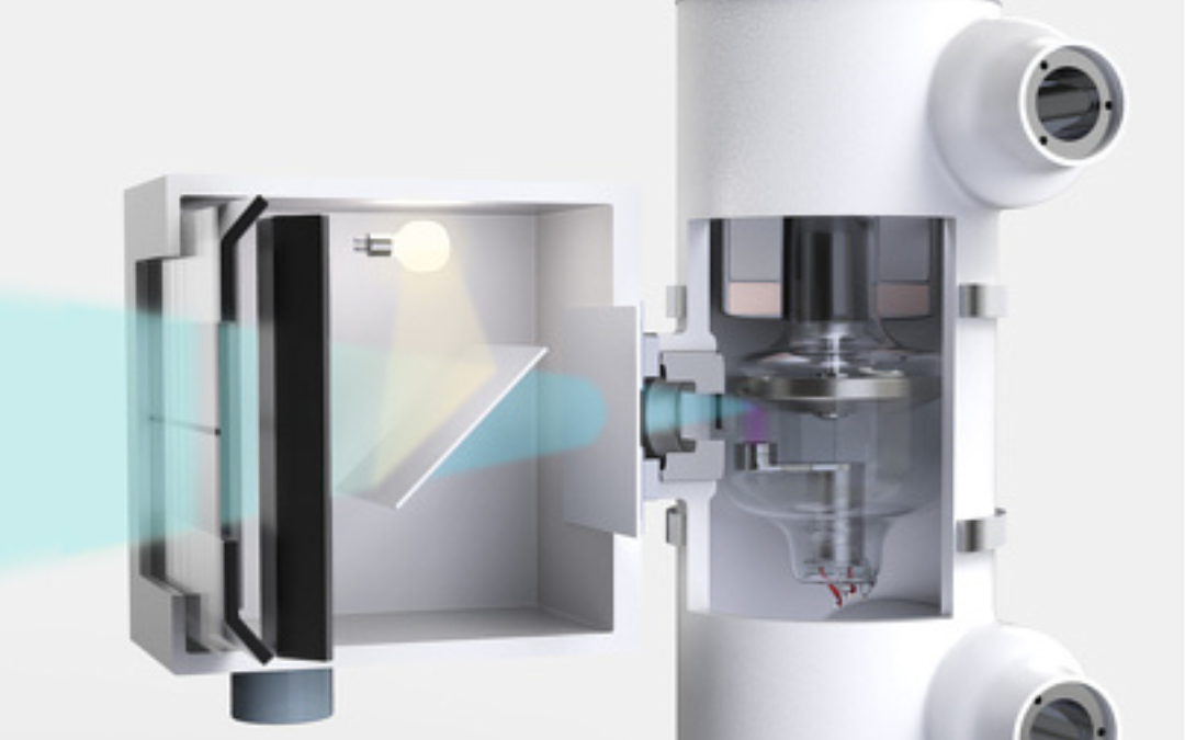
The filter is a metallic sheet which position in the path of x-ray. The process of removing the low energy of x-ray by the help of metallic sheet is called filtration. The x-ray is a heterogeneous photons. The low energy photons make image fog and increase the patient dose but in high energy photons only produce the image. Thus in the path of x-ray the filter are used to remove the low energy photons and reduce the patient dose as well as increase the image quality.
Types Of Filters.
There are two Types of filter.
A) Inherent filtration.
B) Added filtration.
A) Inherent filtration:- The x-ray abortions by the x-ray tube and it’s housing is called inherent filtration. In x-ray tube the anode and cathode are enclosed by the glass envelope and windows of the housing. It is made of aluminum and the thickness of aluminum is 0.5 mm to 1.0 mm.
B) Added filtration:- The added filtration consist of metallic sheet and placed in the path of the primary x-ray beam to absorb low energy photons. The added filter made up of aluminum and copper. The thickness of added filter range 1.0 mm to 1.5 mm . The aluminum is filter low energy photons x-ray because it’s atomic number is Z=13. The copper is best to filter the high energy x-ray because it’s atomic number is Z= 29.

Other types of Filter Are:-
1) Compensating Filter:- Compensating filters are shaped filters used to balance densities on an image when the part difference is large from one end to the other. The compensating filter present in various sizes and shapes. It is made of aluminum but plastic materials also be used. It is used during chest x-ray, foot.
2) Special bow-tie shaped filters:- The special bow-tie shaped filters used in computed tomography imaging system of compensate for the head or body. The shape of filter looks like conic filters either concave or convex . It also used in digital fluoroscopy in which the image intensifier tube is round.
3) Step wedge filter:- It is the adaptation of wedge filters. It is used in intrventional radiology procedures, usually when long section of the anatomy are imaged with the use of two or three separate image receptors. The common application of a step wedge filters involves a three step aluminum wedge and three 14×17″ image receptors for translumber and femoral arteriography and venography.
The filter size and it’s use.
1) in general radiography.
– 1.5 mm aluminum below 70 KVp.
– 2.0 mm Al between 70 to 100 KVp.
– 2.5 mm Al above 100 KVp
2) Mammography.
– Be 1 mm + Mo 0.03 mm (Mo target)
– Be 1 mm + Rh 0.025 mm (Rh target)
Advantage Filter In X-ray.
– The filter remove the low energy photons.
– The filter reduce the patient radiation dose.
– The filter increase the penetration power of x-ray beam.
– The filter increase the image quality.
Why Are Filters Used In X-ray?
Filters are used in X-ray instruments for several reasons:-
A) To reduce patient dose:- X-ray filters can be used to reduce the amount of radiation that a patient is exposed to during an X-ray examination. This helps to minimize the risk of radiation-induced injury or harm to the patient.
B) To improve image quality:- Filters can be used to improve the quality of the X-ray image by removing unwanted radiation and increasing the contrast between different tissue types. This can make it easier for a radiologist to identify and diagnose abnormalities in the image.
C) To increase penetration:- Some filters can be used to increase the penetration of the X-ray beam, allowing for imaging of thicker or denser structures.
D) To reduce scatter radiation:- Scatter radiation, or radiation that is scattered by the patient’s body, can lead to a decrease in image quality and increase patient dose. Filters can be used to reduce scatter radiation and improve image quality.
E) To increase contrast of X-ray images:- Filters can be used to increase the contrast of X-ray images by selectively absorbing certain wavelengths of radiation. For example, Aluminum filter in front of X-ray detector can be used to absorb low energy photons and increase the contrast of bone structures.
F) To reduce noise:-Filters can be used to reduce noise in X-ray images, which can improve image quality and make it easier for a radiologist to identify and diagnose abnormalities.
FAQs.
Q. What is the purpose of filters in an X-ray tube?
Filters are used to selectively remove low-energy X-ray photons, improving image quality and reducing patient radiation dose.
Q. What is inherent filtration in X-ray tubes?
Inherent filtration refers to the filtration provided by the X-ray tube‘s glass envelope and insulating oil, which partially absorbs low-energy X-ray photons.
Q. How does added filtration improve image quality?
Added filtration removes lower-energy X-ray photons, reducing image noise and enhancing the visibility of anatomical details.
Q. What materials are commonly used as X-ray tube filters?
Aluminum is a common material used for added filtration due to its effectiveness in removing low-energy X-ray photons.
Q. Why is beam hardening important in X-ray imaging?
Beam hardening helps optimize the X-ray spectrum for better penetration and image formation, reducing unnecessary patient exposure.
Q. What is a compensating filter and when is it used?
A compensating filter is used to even out X-ray exposure when imaging body parts with varying tissue densities, such as a chest X-ray.
Q. Do filters affect the overall X-ray beam intensity?
Yes, filters reduce the X-ray beam intensity by selectively removing lower-energy photons.
Q. Can improper filter usage affect image quality?
Improper filter usage can lead to suboptimal image quality, increased patient dose, and inadequate diagnostic information.
Q. Are there regulations governing the use of X-ray tube filters?
Yes, regulatory guidelines provide recommendations on the minimum amount of filtration required based on X-ray tube voltage.
Q. How is the thickness of a filter determined?
Filter thickness is determined based on the X-ray tube voltage and the desired level of filtration.
Q. Are filters used only in medical X-ray imaging?
Filters are used in various X-ray applications, including medical, dental, and industrial imaging.
Q. Can excessive filtration lead to underexposed X-ray images?
Excessive filtration can reduce the X-ray beam intensity, potentially leading to underexposed images if exposure settings are not adjusted.
Q. What is half-value layer (HVL) and its relation to filters?
HVL is the thickness of a material that reduces the X-ray intensity by half. Filters are designed to achieve specific HVL values to ensure proper X-ray beam quality.
Q. Do filters affect the energy range of X-ray photons used for imaging?
Yes, filters remove lower-energy X-ray photons, narrowing the energy range and optimizing the X-ray spectrum.
Q. How often should X-ray tube filters be checked for accuracy?
X-ray tube filters should be checked regularly as part of quality assurance and calibration procedures.
Q. Can filters be added or removed easily from an X-ray tube?
Some X-ray systems allow for the addition or removal of filters, while others have fixed filtration.
Q. What is the role of filters in reducing patient radiation dose?
Filters reduce the amount of low-energy radiation reaching the patient, decreasing dose to superficial tissues.
Q. Are there specific filters for pediatric X-ray imaging?
Pediatric imaging may require specialized filters to optimize image quality while minimizing dose.
Q. Do filters affect X-ray tube efficiency or lifespan?
Properly designed filters do not significantly impact X-ray tube efficiency or lifespan.
Q. What is the impact of filters on image contrast?
Filters can improve image contrast by reducing scatter and noise, enhancing the visibility of structures.
Q. Are there any alternatives to aluminum filters?
Some X-ray systems may use other materials, but aluminum is a common and effective choice for added filtration.
Q. How does filter thickness affect X-ray beam quality?
Thicker filters remove more low-energy photons, resulting in higher beam quality.
Q. Can filters be customized for specific imaging needs?
In some cases, filters can be customized to meet specific clinical or imaging requirements.
Q. Do filters impact the overall exposure time for X-ray imaging?
Filters can affect exposure time, as they modify the X-ray beam intensity and energy spectrum.
Q. Can filters correct for overexposed or underexposed X-ray images?
Filters cannot correct exposure errors; proper exposure settings are essential for optimal image quality.
Q. How do filters contribute to patient safety during X-ray procedures?
Filters help reduce unnecessary radiation exposure to patients, enhancing safety.
Q. Are filters part of routine X-ray equipment maintenance?
Yes, filters should be routinely inspected and maintained to ensure proper functioning.
Q. Do filters impact the visibility of soft tissues in X-ray images?
Properly used filters can enhance soft tissue visibility by reducing image noise.
Q. What considerations are important when selecting filters for X-ray imaging?
Factors include the clinical application, X-ray tube voltage, and desired image quality.
Q. How can technologists ensure proper filter usage for optimal X-ray images?
Technologists should follow manufacturer guidelines and quality assurance protocols for filter usage.
FOR MORE CLICK HERE
FOR RADIOLOGY MCQS CLICK HERE
BOOK LINK:- Textbook of Radiology for X-ray, CT, MRI, BSc, BRIT and MSc Technicians

