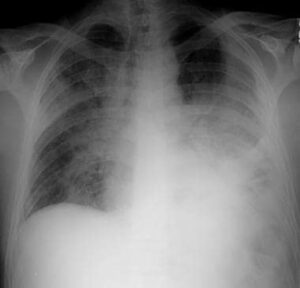
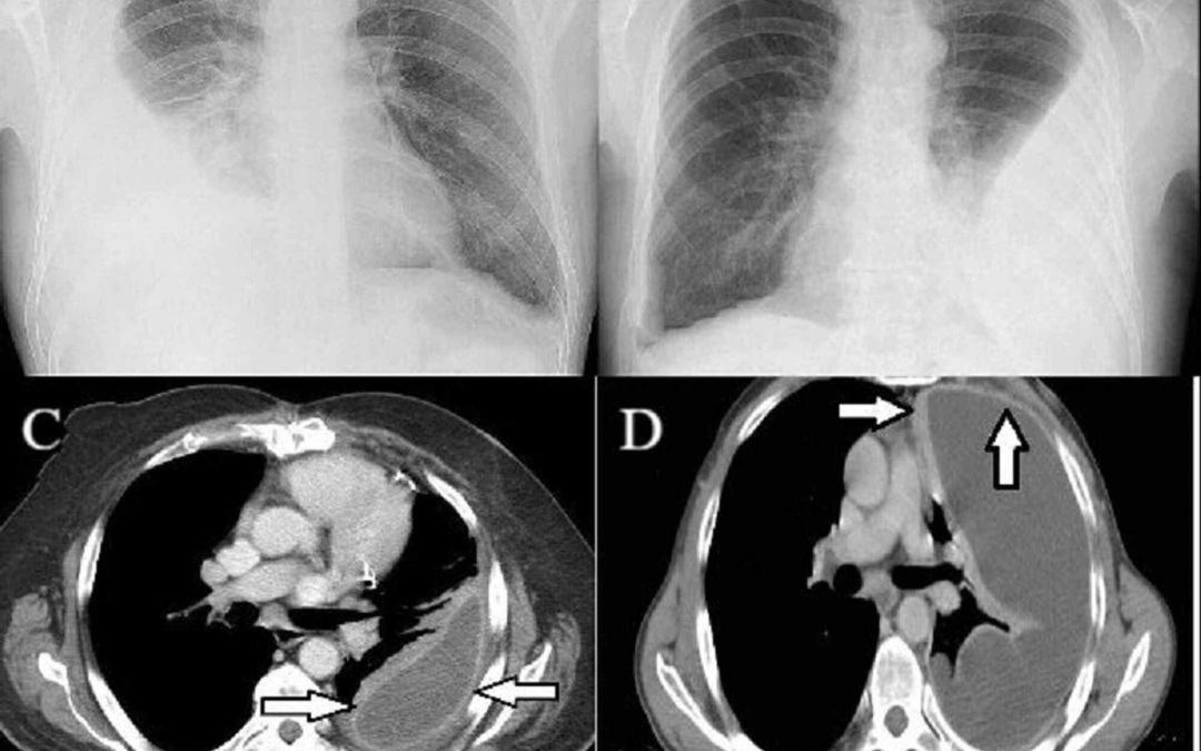
what Is Empyema?
Empyema is a medical condition characterized by the accumulation of pus within a body cavity, often the pleural space between the lungs and chest wall. It is typically a complication of pneumonia or other infections of the respiratory system, and can cause symptoms such as chest pain, coughing, fever, and difficulty breathing.
Empyema can be diagnosed through imaging tests such as chest X-rays or CT scans, and is often treated through drainage of the infected fluid and administration of antibiotics. In some cases, surgery may be necessary to remove the infected tissue and prevent further complications.
Empyema occurs in:-
— The pleural cavity (pleural empyema also known as pyothorax)
— The thoracic cavity
— The uterus (pyometra)
— The appendix (appendicitis)
— The meninges (subdural empyema)
— The joints (septic arthritis)
— The gallbladder
Types Of Empyema.
There are three main types of empyema, classified according to the stage of the disease:
Stage 1: Exudative empyema:- This is the initial stage of empyema where there is an accumulation of fluid in the pleural cavity, but the fluid is not yet infected. This stage is usually treated with antibiotics.
Stage 2 Fibrinopurulent empyema:- In this stage, the fluid in the pleural cavity becomes infected with bacteria, leading to the formation of pus. The pus is often thick and may contain fibrin, a protein that can cause the formation of adhesions within the pleural space. This stage of empyema usually requires drainage of the infected fluid and administration of antibiotics.
Stage 3: Organizing empyema :- This is the most advanced stage of empyema, in which the pus becomes thick and forms a thick layer of scar tissue. This scar tissue can cause the pleural space to become smaller, making it difficult for the lungs to expand fully. This stage of empyema may require surgery to remove the scar tissue and re-expand the lung.
What Are The Symptoms Of Empyema?
The symptoms of empyema may vary depending on the severity and stage of the disease, but common symptoms include:-
1. Chest pain, which may be sharp or dull
2. Cough, which may be dry or produce phlegm
3. Shortness of breath or difficulty breathing
4. Fever, which may be high
5.Chills and sweats
6.Fatigue or weakness
7.Loss of appetite and weight loss
8. Rapid heart rate
In severe cases, empyema can cause complications such as sepsis, lung abscesses, and respiratory failure, which can be life-threatening. Therefore, it is important to seek medical attention if you experience any of these symptoms.
What Causes Empyema?
Empyema is usually caused by a bacterial infection in the lungs or respiratory system. The most common cause of empyema is pneumonia, which is an infection of the lung tissue. Bacteria from the pneumonia infection can spread to the pleural space, causing inflammation and the accumulation of pus.
Other conditions that can cause empyema include lung abscesses, tuberculosis, bronchial obstruction, and chest trauma. In some cases, empyema may also occur as a complication of chest surgery or as a result of an infection in another part of the body that spreads to the pleural space.
Risk factors for empyema include smoking, alcohol abuse, a weakened immune system, and underlying respiratory or lung conditions. It is important to treat respiratory infections promptly to reduce the risk of developing empyema.
What Tests Will Be Done To Diagnose Empyema?
The diagnosis of empyema typically involves a combination of physical examination, imaging tests, and laboratory tests.
1. Physical examination:- The doctor will listen to your chest with a stethoscope to check for abnormal breathing sounds and may tap your chest to check for areas of tenderness or fluid buildup.
2. Imaging tests:- Chest X-rays and CT scans can help detect the presence of fluid or pus in the pleural space and determine the extent of the infection. Ultrasound may also be used to guide the placement of a drainage tube.


3. Laboratory tests:- Samples of the pleural fluid are taken and analyzed to determine the type of bacteria causing the infection and guide the selection of antibiotics. Blood tests may also be done to check for signs of infection.
In some cases, a pleural biopsy may be performed to obtain a sample of the pleural tissue for analysis. The results of these tests can help confirm the diagnosis of empyema and guide the appropriate treatment.
Treatment Of Empyema.
The treatment of empyema typically involves draining the infected fluid from the pleural space and administering antibiotics to control the infection. In some cases, surgery may be necessary to remove infected tissue or prevent the formation of scar tissue.
1. Drainage:- A chest tube is usually inserted into the pleural space to drain the infected fluid. The tube is left in place until the fluid stops accumulating and the infection is under control. The tube may be connected to a suction device to help drain the fluid more quickly.
2. Antibiotics:- Antibiotics are typically given to treat the bacterial infection causing the empyema. The choice of antibiotic depends on the type of bacteria causing the infection and the results of lab tests.
3. Surgery:- If the empyema does not respond to drainage and antibiotics, surgery may be necessary. The type of surgery depends on the stage and severity of the empyema but may include decortication, which involves removing the infected tissue and scar tissue from the pleural space.
In addition to medical treatment, supportive care may be necessary to manage symptoms and prevent complications. This may include pain management, oxygen therapy, and respiratory support.
It is important to treat empyema promptly to prevent complications and improve outcomes.
BOOK LINK :-https://amzn.to/3MDM8KP
Radiographic Pathology (Point (Lippincott Williams & Wilkins)
Also Read 1. https://www.radiologystar.com/bouveret-syndrome/
2.https://www.radiologystar.com/kartangeners-syndrome/
3, https://www.radiologystar.com/duodenal-adenocarcinoma/


This is nice package for every imaging student