What Is Dacrocystography?
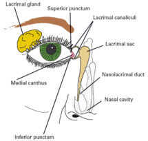
Dacrocystography is the radiolological examination to help the study of nasolacrimal system by the injection of contrast media through cannaliculi of the eyelid.
During Dacrocystography, a contrast agent is injected into the tear ducts through a small tube or catheter that is inserted into the puncta, which are tiny openings located at the inner corner of the eye.

Indication.
— Epiphora.
— Obstruction of nasolacrimal duct.
— Chronic dacrocystisis.
— Fistula.
–Tumors.
— Diverticula.
–Dacrolith.
Contraindications.
— Severe infection in lacrimal duct.
— Hypersensitivity to contrast media.
Contrast Media.
— Lipiodol
— Dose :- 0.5-2ml.
Equipment.
— Fluoroscopy unit.
— Spot film device.
— 2cc Syringe.
— Lacrimal cannula.
— Local anesthesia.
— Puncture dilator
— Cotton.
Patient Preparation.
— None.
Preliminary film.
— Lateral view of skull.
— Occipito-mental view.
— AP view.
— Oblique view.
Procedure / Technique.
— The patient lies on supine position on the fluoroscopy table.
— Clean the patient’s eye with normal saline water for prevention of any infection.
— The local anesthesia is applied in eye to make patient relaxed.
— Then the lacrimal puncture dilator are applied for dilation of lacrimal duct.
— When the lacrimal then the Radiologist located lower canaliculus at the middle end of lid and puncture the duct through lacrimal cannula.
— Then around 1 – 2ml of contrast injected to opacify the entire nasolacrimal apparatus under the fluoroscopy observation.
— Than take a spot film
— After end of the examination the cannula is removed.
Filming.
— AP view.
— Lateral.
— Oblique view.
— Occipito-mental view.


Complications.
— Contrast extravasation.
— Granuloma formation.
— Pain .
— Bad test in mouth.
— Infection.
Aftercare.
– To keep the patient on observation of 1-2 hrs to show the any types of difficulty to patient like eaching, burning and redness of eye.
Types Of Dacrocystography.
There are several way of types to perform Dacrocystography.
A) Conventional Dacrocystography
B) Distension Dacrocystography.
C) Macrography Dacrocystography.
D) Seriography Dacrocystography.
E) Digital Subtraction Dacrocystography
E) Kinetic conventional Dacrocystography.
F) Real-time Dacrocystography.
G) Computed Tomography Dacrocystography.
H) Magnetic Resonance Dacrocystography .
FAQs.
Q. Which method is used for Dacryocystography?
Conventional cannulation dacryocystography is invasive and requires administration of local anaesthesia, cannulation, dilatation of the lacrimal punctum, and injection of contrast material.
Q. What is Dacryocystitis?
Dacryocystitis is inflammation of the lacrimal sac
Q. What is Dacryocystography an investigation for evaluation of?
Dacryocystography, one of the imaging methods that enable us to investigate the anatomy of the lacrimal duct, is mainly used in patients with epiphora.
Q. What are the advantages of CT DCG?
CT-DCG is an excellent tool for delineating the bony structures around the lacrimal system and to some extent soft tissue study of lacrimal system
Q. Is Dacryocystography still performed today?
No It’s done today.
FOR MORE CLICK HERE
BOOK LINK :- Fundamentals of Special Radiographic Procedures

