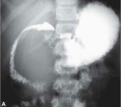
What Is Barium Meal?
The Barium meal is the radiological examination to help the study of the Stomach, duodenum and proximal jejunum by injected of barium Sulfate contrast media through orally.
Indication Of Barium Meal.
– Abdominal mass.
— Dysplasia
— Weight loss
— Gastrointestinal hemorrhage
— Obstruction
— Carcinoma
— Abdominal pain
— Malformation
— Vomiting
— Anemia
— Tuberculosis
— Gastro-oessophageal reflux
— Heart burn
— Anorexia
— Polyps
— To rule out perforation
Contraindications Of Barium Meal.
— Suspected perforation.
— Completely bowel obstruction.
— Suspected Fistula.
— Recent biopsy or abdominal surgery.
Contrast Media For Barium Meal.
— 100% W/V barium sulfate suspension for single contrast method.
— 250% W/V Barium sulfate for double contrast method.
Equipment.
— Fluoroscopy unit
— Spot film device
— Barium pot
Patient preparation For Barium Meal.
— The patient must be NPO 5 – 6 Hrs before the examination.
— The patient must be give the instructions to take low diet for one to two day before the examination.
— Laxative may be given to patient to remove the all feces material from the abdomen.
— The patient must be stop smoking or tobacco for prevent the proper coating of barium sulfate on the mucosal.
Procedure / Technique For Barium Meal .
— When the patient come in radiology department.
— To ask the patient to remove all the clothes and wear the hospital gown.
— To explain the whole Procedure before the patient.
— Take the consent from to patient or patient attender.
This procedure done by two methods.
A) Single contrast method.
B) Double contrast method.
A) Single contrast method.
— The patient lies on supine position in fluoroscopy table.
–The barium suspension 80-100% W/V solution used in this method.
— The solution of barium sulfate is introducing the patient mouth and swallow under the observation of Fluoroscopy unit.
— When the patient is swallow 3-4 mouth full of barium sulfate solution .
— Then the rotate the patient in clock wise for coating barium on the stomach mucosa under observation in fluoroscopy.
— Then kept the patient in prone position to enter the barium sulfate solution in the duodenum.
— Than take the Right anterior oblique position film.
— AP film taken for duodenum.
B) Double contrast method.
The double contrast method, help the study of mucosal details in abdomen
— The patient lies on supine position in fluoroscopy table.
— The high density barium sulfate solution (250% W/v) used.
— The Buscopan 2ml IV injection injected in the patient body.
— The barium sulfate solution is giving in to the patient mouth and give instructions to drink the solution.
— When the patient is drinking 3-4 full mouth .
— Then rotate the patient in clock wise to coated the barium sulfate solution in stomach mucosal in observation in fluoroscopy.
— The the patient is in prone position to enter the barium sulfate solution in duodenum.
— Then take a film.
Filming.
A) for single contrast method.
— AP in supine:- For Fundus of stomach
— Prone or supine:- For body and antrum.
— RAO :- For greater curvature.
— LAO :- Lesser curvature.
— RAO :- Duodenum cup and loop.
B) Double contrast method.
— Erect :- for Fundus
— Supine :- for Body and antrum
— LAO:- for lesser curvature
— RAO :- for Greater curvature
— LAO :- for duodenum cup and loop
Aftercare.
— Ask the patient to take a maximum fluid.
— To inform the patient about feces will be white for some days.
— Gave the Dulcolax to remove the barium sulfate solution through the feces.
Complications.
— Leakage barium sulfate solution for perforation.
— Aspiration.
— It may caused appendicitis.
Also Read:- Barium Swallow
BOOK LINK :- A Guide to Radiological Procedures Paperback
Clark’s Procedures in Diagnostic Imaging: A System-Based Approach

