What Is 11 Week Ultrasound ?
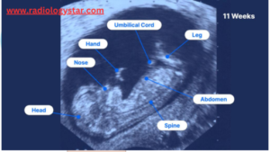
An 11 week ultrasound, also known as a dating scan or a first-trimester ultrasound. 11 week ultrasound is a medical procedure performed during pregnancy to assess the development and well-being of the fetus. This ultrasound is typically done around the 11th week of gestation, which is roughly the end of the first trimester.
It’s important to note that the 11 week ultrasound is not as detailed as the anatomy scan (usually done around 18-20 weeks) when more comprehensive assessments of fetal anatomy are conducted. The primary goal of the 11 week ultrasound is to assess the basic health and development of the fetus and to provide important information for the remainder of the pregnancy.
Here are some of the primary purposes and things that can be observed during an 11 week ultrasound:-
A. Confirming Pregnancy:- The ultrasound confirms the presence of a viable pregnancy and helps determine the gestational age, which is crucial for estimating the due date.
B. Fetal Size and Growth:- The ultrasound measures the size of the fetus, which can help determine if the pregnancy is progressing as expected based on the estimated due date.
C. Detecting Multiple Pregnancies:- An 11 week ultrasound can reveal if there is more than one fetus (twins, triplets, etc.).
D. Checking for Major Anomalies:- While it’s not as comprehensive as the more detailed anatomy scan done later in pregnancy, the 11 week ultrasound may detect some major fetal anomalies or issues with development.
E. Nuchal Translucency Measurement:- One of the key assessments during an 11 week ultrasound is the measurement of nuchal translucency. This is the thickness of the fluid-filled space at the back of the baby’s neck. Abnormalities in this measurement can be associated with certain chromosomal abnormalities, such as Down syndrome.
F. Checking the Placenta and Amniotic Fluid:- The ultrasound can also assess the placement and health of the placenta and the amount of amniotic fluid in the uterus.
G. Listening to the Fetal Heartbeat:- In some cases, the ultrasound may allow the healthcare provider to listen to the fetal heartbeat, which is a reassuring sign of a healthy pregnancy.
Baby’s Organs at an 11 Week Ultrasound.
During an 11 week ultrasound, the development of the baby’s organs is in progress, but many of the organs are still forming and may not yet be fully developed. At this stage of pregnancy, the focus is on assessing the basic development and health of the fetus, rather than detailed organ examination.
Here’s an overview of the state of some of the baby’s organs and systems at 11 weeks of gestation:-
A. Brain:- The brain has begun forming, and the basic structures are present. However, it’s still in the early stages of development.

B. Heart:- The heart is rapidly developing and should be visible on the ultrasound. The four chambers of the heart are forming, and the heartbeat can often be detected and measured during the 11-week ultrasound.
C. Lungs:- The lungs are in the early stages of development and consist mainly of tiny air sacs called bronchioles.
D. Liver:- The liver is present and functioning in basic capacities, but it is still developing.
E. Kidneys:- The kidneys are forming and are visible on the ultrasound. They will continue to develop and mature throughout the pregnancy.
F. Digestive System:- The basic structures of the digestive system, including the stomach and intestines, are forming.
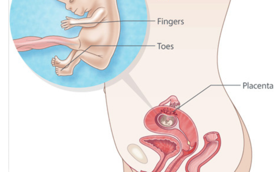
G. Limbs:- The arms and legs are present and will continue to grow and develop throughout the pregnancy. Fingers and toes may be seen, but they are still in the early stages of formation.
H. Genitalia:- The gender of the fetus may be determined by the presence or absence of certain genital structures, but it may not always be clearly visible at this stage.
I. Spine:- The spine is visible on the ultrasound, and the basic vertebrae are forming.
11 Week Fetal Body Shape And Size Measurement.
During an 11 week fetal ultrasound, the baby’s body shape and size are still in the early stages of development. However, some basic measurements and observations can be made to assess the growth and progress of the fetus.
Here are some key points related to the fetal body shape and size measurement at 11 weeks:-
A. Crown-Rump Length (CRL):- The primary measurement taken during an 11 week ultrasound is the crown-rump length. This measurement involves determining the length of the fetus from the crown (the top of the head) to the rump (the bottom of the spine). CRL is used to estimate the gestational age and is a key factor in determining the due date. At 11 weeks, the average CRL is approximately 4.1 to 5.1 cm (about 1.6 to 2 inches).
B. Body Proportions:- At 11 weeks, the fetus has a relatively large head compared to the rest of its body. This is a normal characteristic of early fetal development, and as the pregnancy progresses, the body will catch up in size, resulting in a more proportionate appearance.
C. Limb Development:- Limb buds (arms and legs) are present and beginning to develop. Fingers and toes may be in the early stages of formation, but they are not fully developed or separated yet.
D. Body Flexion:- The fetus is still in a relatively curled or flexed position within the uterus. This is due to the limited space available at this stage of development.
E. Amniotic Fluid:- The fetus is surrounded by amniotic fluid, which provides protection and cushioning. The amount of amniotic fluid is assessed during the ultrasound to ensure it is at a healthy level for fetal development.
F. Overall Appearance:- The fetus may appear somewhat translucent on the ultrasound, and its body contours may be less defined than in later stages of pregnancy. Organs and features are still developing and may not be fully visible or distinguishable at this stage.
11 Week Fetal Baby Body Development
During the 11th week of pregnancy, fetal development is progressing rapidly, and the baby is undergoing significant changes in terms of body development.
Here’s an overview of what’s happening during the 11th week of gestation:-
A. Crown-Rump Length (CRL):- At 11 weeks, the fetus measures approximately 4.1 to 5.1 centimeters (about 1.6 to 2 inches) in crown-rump length. This measurement is taken from the top of the baby’s head (crown) to the bottom of the spine (rump) and is used to estimate gestational age.
B. Head Development:- The baby’s head is relatively large compared to the rest of its body at this stage. The brain is developing rapidly, and the facial features are becoming more defined. The eyes, nose, and mouth are present but may still appear quite large in proportion to the head.
C. Limb Development:- The baby’s arms and legs are growing and becoming more distinct. Fingers and toes are forming and may be separated by tiny webs of skin.
D. Body Flexion:- The fetus is still in a curled or flexed position within the uterus due to the limited space. This fetal position will gradually change as the baby grows and develops.
E. Facial Features:- The baby’s facial features are developing, including the formation of eyelids and external ears. However, these features are not fully developed or functional at this stage.
F. Genitalia:- By the 11th week, the baby’s external genitalia may have started to develop, but it may not be possible to determine the baby’s gender via ultrasound with certainty. The genitals will continue to mature throughout the pregnancy.
G. Digestive System:- The basic structures of the digestive system, including the stomach and intestines, are forming. The baby swallows amniotic fluid, and some early digestive functions are initiated.
H. Cardiovascular System:- The heart has already formed and is beating. Blood is circulating throughout the body, and the heart is pumping blood to supply oxygen and nutrients to the developing organs and tissues.
I. Muscle and Bone Development:- The baby’s muscles and bones are developing, but they are still quite soft and flexible at this stage.
J. Amniotic Fluid:- The baby is surrounded by amniotic fluid, which provides protection and cushioning. The amount of amniotic fluid is monitored to ensure it remains at a healthy level.
Reasons To Get An Ultrasound At 11 Weeks Pregnant
An ultrasound at 11 weeks of pregnancy, often referred to as a first-trimester ultrasound or dating scan, serves several important purposes:
A. Confirm Pregnancy:- An 11 week ultrasound can confirm the presence of a viable pregnancy, ensuring that the fertilized egg has successfully implanted in the uterus and that fetal development is underway.
B. Estimate Gestational Age:- The ultrasound measures the crown-rump length (CRL) of the fetus, which helps estimate the gestational age. This estimation is crucial for determining the due date, which is important for scheduling prenatal care and monitoring the progress of the pregnancy.
C. Detect Multiple Pregnancies:- Ultrasound can identify whether there is more than one fetus (e.g., twins, triplets), as multiple pregnancies require specialized care and monitoring.
D. Nuchal Translucency Measurement:- During the 11 week ultrasound, the thickness of the nuchal translucency (the fluid-filled space at the back of the baby’s neck) is measured. Abnormalities in this measurement may be associated with chromosomal conditions like Down syndrome. This measurement can help assess the risk of certain genetic disorders.
E. Check for Major Anomalies:- While not as comprehensive as later anatomical scans, an 11 week ultrasound can identify some major fetal abnormalities, helping parents make informed decisions about their pregnancy.
F. Monitor Basic Fetal Development:- The ultrasound may show the development of key structures, such as the fetal heart, limbs, and organs, to ensure they are forming as expected.
G. Placental Location:- It can determine the location of the placenta, which is important for monitoring its health and ensuring it’s not covering the cervix (placenta previa).
H. Assess Amniotic Fluid:- The amount of amniotic fluid surrounding the fetus is assessed to ensure it is at a healthy level for fetal development.
I. Reassurance:- Seeing and hearing the fetal heartbeat during this ultrasound can provide emotional reassurance to the expectant parents, confirming that the pregnancy is progressing as expected.
J. Early Intervention:- In cases where abnormalities or concerns are identified during the 11 week ultrasound, it can lead to early interventions, additional testing, or consultations with specialists to address any issues or provide guidance for the pregnancy.
FAQs.
Q. What is the purpose of an 11-week ultrasound?
The primary purpose is to assess fetal development, estimate gestational age, and screen for certain conditions like Down syndrome.
Q. Is the 11-week ultrasound a routine part of prenatal care?
It may be recommended as part of routine prenatal care, but it’s not always necessary for every pregnancy.
Q. How is the gestational age of the fetus determined during this ultrasound?
The ultrasound measures the crown-rump length (CRL) of the fetus, which helps estimate gestational age.
Q. Can I find out the gender of the baby at 11 weeks?
It’s usually too early to determine the baby’s gender accurately at this stage.
Q. What is nuchal translucency, and why is it measured during this ultrasound?
Nuchal translucency is the thickness of the fluid-filled space at the back of the baby’s neck. It’s measured to assess the risk of certain chromosomal abnormalities, like Down syndrome.
Q. How is the nuchal translucency measurement taken?
It’s measured using ultrasound by identifying the clear space at the back of the baby’s neck.
Q. What does an elevated nuchal translucency measurement indicate?
An elevated measurement may indicate an increased risk of chromosomal abnormalities, but it doesn’t provide a diagnosis.
Q. How accurate is the risk assessment for chromosomal abnormalities based on this ultrasound?
It provides an estimate of risk but is not a definitive diagnostic test.
Q. Are there any risks associated with the ultrasound procedure?
Ultrasound is generally considered safe, with no known harmful effects when used appropriately.
Q. Can I hear the baby’s heartbeat during the 11 week ultrasound?
Yes, in many cases, you can hear the fetal heartbeat, which is reassuring.
Q. What can be seen in terms of the baby’s body development?
Basic organ formation, limb development, and overall growth can be observed.
Q. Is it possible to identify any major anomalies during this ultrasound?
While some major anomalies may be detectable, a more detailed anatomy scan is typically performed later in pregnancy.
Q. Can I see the baby’s facial features on the ultrasound?
Facial features are forming, but they may not be fully distinguishable at this stage.
Q. What is the typical fetal position at 11 weeks?
The fetus is usually in a curled or flexed position due to limited space in the uterus.
Q. What information can be gained about the amount of amniotic fluid?
The ultrasound assesses the amount of amniotic fluid to ensure it is at a healthy level for fetal development.
BOOK LINK :- Examination Review for Ultrasound: Abdomen and Obstetrics & Gynecology
BOOK LINK:- Sonography


