

What Is 7 Weeks Ultrasound ?
A 7 weeks ultrasound is a medical imaging procedure that uses high-frequency sound waves to create images of a developing fetus inside the womb. This type of ultrasound is typically performed early in a pregnancy to confirm the gestational age of the fetus, check for the presence of a heartbeat, and detect any abnormalities.
During a 7 weeks ultrasound, the healthcare provider will use a handheld device called a transducer to emit sound waves into the abdomen. These waves bounce off the developing fetus and create images that can be viewed on a monitor. The procedure is non-invasive and typically does not cause any discomfort or harm to the mother or the developing fetus.
A 7 weeks ultrasound, also known as a first trimester ultrasound, is an ultrasound exam performed during the early stages of pregnancy. At 7 weeks pregnant, this ultrasound is usually done for the following purposes:-
A) To confirm the pregnancy:- In 7 weeks ultrasound can confirm that a pregnancy exists and that the embryo is developing inside the uterus. It can also detect the baby’s heartbeat, which confirms that the pregnancy is viable.
B) To determine the cause of any bleeding:- 7 weeks ultrasound can check if the bleeding is from the uterine implantation site or if there is any other issue. It can also check if the baby is developing normally despite the bleeding.
C) To determine the due date:- In 7 weeks ultrasound can accurately determine the gestational age of the baby and provide an expected due date for delivery.
D) To check for multiples:- In 7 weeks ultrasound can determine if you are carrying twins or triplets by detecting the number of embryos and heartbeats.
E) To look for any abnormalities:- In 7 weeks ultrasound may detect some major abnormalities that could affect the viability or development of the pregnancy. The ultrasound at this stage however cannot detect all abnormalities accurately. Further advanced ultrasounds are required for a more comprehensive assessment.
Measurement Of 7 Weeks Ultrasound?
The measurements of 7 weeks ultrasound provide important information about the viability and normal development of an early pregnancy. If the measurements are off or no embryo with a heartbeat is detected at 7 weeks, it could indicate a non-viable pregnancy or other issues that need further assessment. Repeat ultrasound scans may be required in some cases.
During a 7 weeks ultrasound, the following measurements are typically taken to assess the development of the embryo:-
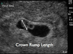
A) Crown-rump length (CRL):- This measures the length of the embryo from the top of the head (crown) to the bottom of the buttocks (rump). At 7 weeks, the CRL is usually around 5-9 mm. This measurement is very accurate for dating the pregnancy at this stage.
B) Heart rate:- The embryo’s heart starts beating at around 6 weeks. At a 7 weeks ultrasound, the heart rate is usually between 120-170 beats per minute. A normal heart rate indicates that the pregnancy is progressing well so far.

C) Yolk sac:- The yolk sac provides nutrients to the embryo until the placenta develops. At 7 weeks ultrasound, a yolk sac measuring around 3-5 mm is usually seen. The yolk sac will eventually disappear by week 10 as the placenta takes over.

D) Gestational sac:- The gestational sac encloses the embryo and yolk sac. At 7 weeks ultrasound, the average diameter of a normal gestational sac is around 18-24 mm. The sac will continue to grow as the pregnancy progresses.
E) Fetal pole:- The fetal pole is the thickening on the margin of the yolk sac where the embryo develops. At 7 weeks, a fetal pole of around 3-5 mm in length indicates a normal developing embryo.
What Are The Possible Reasons For Non-viable Pregnancy At 7 Weeks?
A non-viable pregnancy at 7 weeks ultrasound may refer to a pregnancy that has stopped developing or is unlikely to result in a live birth. non-viable pregnancy occur due to chromosomal or other abnormalities that do not indicate a lasting fertility issue.
Some possible reasons for a non-viable pregnancy detected at a 7 week ultrasound include:-
A) Miscarriage:- The most common reason for a non-viable pregnancy at 7 weeks is miscarriage or early pregnancy loss. If there is no embryo with a heartbeat seen on ultrasound at 7 weeks, or if the embryo measures significantly smaller than expected with no heartbeat, it likely indicates a miscarriage.
B) Ectopic pregnancy:- An ectopic pregnancy is when the embryo implants outside the uterus, usually in the fallopian tubes. At 7 weeks, an ectopic pregnancy may show an abnormal looking gestational sac or no clear gestational sac in the uterus. There may also be no embryo with heartbeat detected. An ectopic pregnancy requires immediate medical treatment.
C)Molar pregnancy:- A molar pregnancy occurs when a non-viable egg is fertilized and there is abnormal placental tissue growth. On a 7 week ultrasound, there may be no embryo detected or an atypically large placental mass with no embryo. HCG levels are also usually very high in a molar pregnancy. It requires treatment to remove the placental tissues.
D) Blighted ovum:- A blighted ovum refers to a fertilized egg that fails to develop into an embryo. There is a gestational sac but no embryo and no yolk sac or heartbeat detected at 7 weeks. It eventually ends in a miscarriage.
E) Inaccurate dating:- Sometimes a pregnancy may be not as advanced as first thought based on the last menstrual period. This can lead to no embryo or heartbeat being detected at a 7 week ultrasound. Follow up scans may determine a more accurate due date and development of the pregnancy.
F) Chromosomal abnormalities:- Chromosomal abnormalities are the most common cause of early pregnancy loss. They occur when there is a problem with the genetic material of the developing fetus, which can prevent it from developing normally.
G) Infection:- Infections can cause early pregnancy loss by damaging the developing fetus or interfering with the implantation process. Common infections that can lead to pregnancy loss include rubella, toxoplasmosis, and sexually transmitted infections (STIs).
H) Hormonal imbalances:- Hormonal imbalances, such as low levels of progesterone, can interfere with the development of the uterine lining and prevent the embryo from implanting or growing properly.
What Are The Next Steps If A Non-viable Pregnancy Is Suspected?
If a non-viable pregnancy is suspected, the next steps will depend on the individual circumstances and the cause of the suspected pregnancy loss. Here are some possible next steps that a healthcare provider may recommend:
A) Repeat ultrasound:- If a non-viable pregnancy is suspected based on an initial ultrasound, the healthcare provider may recommend a repeat ultrasound in a few days or a week to confirm the diagnosis.
B) Blood tests:- Blood tests can measure the levels of pregnancy hormones in the blood, such as human chorionic gonadotropin (HCG) and progesterone. If the levels of these hormones are not consistent with a viable pregnancy, it may indicate a non-viable pregnancy.
C) Treatment options:- If a non-viable pregnancy is confirmed, the healthcare provider will discuss treatment options with the individual. Depending on the circumstances, options may include expectant management, medication to induce miscarriage, or a procedure to remove the pregnancy tissue.
D) Follow-up care:- After a non-viable pregnancy, follow-up care is important to monitor for any complications and ensure that the individual’s physical and emotional health needs are addressed
What Are The Chances Of A Successful Pregnancy After A Non-viable One?
The chances of a successful pregnancy after a non-viable pregnancy depend on several factors, including the cause of the non-viable pregnancy and the individual’s health and medical history. Here are some factors that can affect the chances of a successful pregnancy after a non-viable one:-
A) Age:- Age can be a significant factor in pregnancy outcomes. Women who are older may have a higher risk of pregnancy loss and a lower chance of a successful pregnancy after a non-viable one.
B) Cause of pregnancy loss:- The cause of the non-viable pregnancy can also affect the chances of a successful pregnancy. If the cause was a chromosomal abnormality, for example, the risk of a subsequent pregnancy with a chromosomal abnormality may be higher.
C) Health history:- A history of previous pregnancy loss, medical conditions such as thyroid disorders, and lifestyle factors such as smoking or substance use can also affect the chances of a successful pregnancy.
D) Treatment:- The type of treatment received for the non-viable pregnancy can also affect the chances of a successful pregnancy.
What Are The Treatment Options For A Non-viable Pregnancy?
The treatment options for a non-viable pregnancy will depend on the individual circumstances and the stage of pregnancy. Here are some possible treatment options:-
A) Expectant management:- In some cases, a non-viable pregnancy will pass naturally without medical intervention. This is known as expectant management or “watchful waiting.” During this time, the healthcare provider will monitor the individual’s condition to ensure that there are no complications.
B) Medication:- Medications may be prescribed to help induce the miscarriage if the pregnancy is not passing on its own. The most common medication used is misoprostol, which helps to soften and open the cervix to allow the pregnancy tissue to pass. This medication may be administered vaginally or orally, and the individual will typically experience cramping and bleeding similar to a heavy period.
C) Surgical options:- If the pregnancy does not pass with expectant management or medication, a surgical procedure may be necessary. The two main types of surgical procedures are dilation and curettage (D&C) and suction dilation and curettage (S&D). These procedures involve removing the pregnancy tissue from the uterus under general anesthesia.
D) Watchful waiting:- If the individual has an ectopic pregnancy, the healthcare provider may recommend watchful waiting to see if the pregnancy resolves on its own. If the ectopic pregnancy is not resolving or is causing significant symptoms or complications, surgery may be necessary to remove the pregnancy.
BOOK LINK:-https://amzn.to/3LyA1fU
Sonography
BOOK LINK :-https://amzn.to/44xe9Ky
Craig’s Essentials of Sonography and Patient Care 4th Edition