
What Is 3D Ultrasound ?
What Is The Purpose Of a 3D Ultrasound?
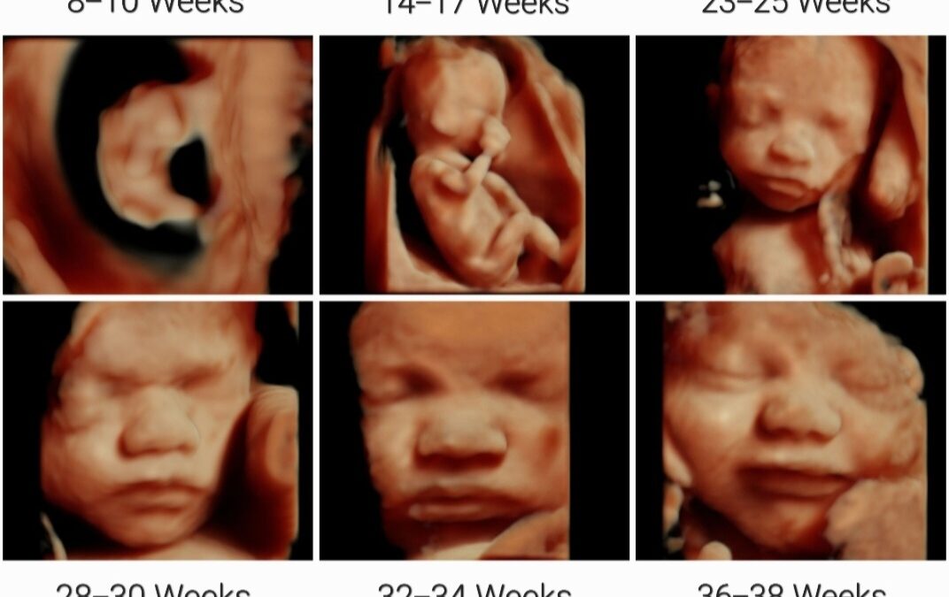
3D ultrasounds can help doctors and expectant parents to better understand the fetal anatomy and detect any abnormalities or developmental issues. They can also provide a more vivid and realistic image of the fetus, which can be helpful for bonding and emotional support for parents. In addition to providing more detailed images of the fetus, 3D ultrasounds can also be used for diagnostic purposes, such as checking the growth and development of the fetus and monitoring for any complications during pregnancy.
What Are The Use Of 3D Ultrasound ?
Advantage of 3D Ultrasound.
FAQs:-
Q. What is a 3D ultrasound?
A 3D ultrasound is a medical imaging technique that uses sound waves to create three-dimensional images of the fetus in the womb or other body structures.
Q. How is a 3D ultrasound different from a 2D ultrasound?
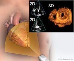
A 2D ultrasound provides flat, two-dimensional images, while a 3D ultrasound generates more detailed, three-dimensional images.
Q. When is a 3D ultrasound typically performed during pregnancy?
3D ultrasounds are usually performed between 24 and 32 weeks of pregnancy to capture clearer images of the developing fetus.
Q. Is a 3D ultrasound safe for the baby?
Yes, 3D ultrasounds are considered safe for both the mother and the baby as they use sound waves and do not involve ionizing radiation.
Q. Why might a doctor recommend a 3D ultrasound?
A doctor may recommend a 3D ultrasound to get a better view of fetal development or to assess specific medical conditions.
Q. How long does a 3D ultrasound procedure usually take?
The duration of a 3D ultrasound can vary but typically lasts between 15 to 30 minutes.
Q. Can I determine the gender of the baby through a 3D ultrasound?
In some cases, a 3D ultrasound can reveal the gender of the baby, but it is not always guaranteed.
Q. Are 3D ultrasounds covered by insurance?
Coverage for 3D ultrasounds varies depending on the insurance provider and the medical necessity of the procedure.
Q. Are there any preparation requirements for a 3D ultrasound?
Preparation requirements may vary, but generally, a full bladder is not necessary for 3D ultrasounds.
Q. Can I request a 3D ultrasound if it’s not medically necessary?
Some healthcare facilities may offer elective 3D ultrasounds for non-medical purposes upon request.
Q. How much does a 3D ultrasound typically cost?
The cost of a 3D ultrasound can vary based on the healthcare provider and location. It may range from $100 to $500 or more.
Q. What are the benefits of 3D ultrasound in prenatal care?
3D ultrasound can provide clearer images, aiding in the early detection of certain birth defects or abnormalities.
Q. Are there any risks associated with 3D ultrasounds?
3D ultrasounds are considered safe, but they should only be performed by qualified medical professionals to minimize any potential risks.
Q. Can 3D ultrasound detect chromosomal abnormalities?
3D ultrasounds can provide additional information, but they are not the primary method for diagnosing chromosomal abnormalities.
Q. How should I prepare for a 3D ultrasound appointment?
Follow any specific instructions provided by your healthcare provider, such as drinking water before the appointment.
Q. Can I get a 3D ultrasound if I’m having twins or multiples?
Yes, 3D ultrasounds can be performed for pregnancies with twins or multiples.
Q. Are there limitations to what a 3D ultrasound can show?
While 3D ultrasounds offer more detail, they may not capture every aspect of fetal development or certain medical conditions.
Q. Can a 3D ultrasound be performed at home using mobile apps or devices?
It is not recommended to rely on mobile apps or devices for medical imaging purposes, including 3D ultrasound.
Q. What is the difference between a 3D and 4D ultrasound?
A 3D ultrasound provides static three-dimensional images, while a 4D ultrasound adds the element of time, creating a real-time video effect.
Q. Can 3D ultrasounds be used for other medical imaging purposes besides pregnancy?
Yes, 3D ultrasound technology is used in various medical fields, including cardiology and gynecology, for diagnostic purposes.
Q. Are 3D ultrasounds used in non-medical settings, such as entertainment?
Some non-medical facilities may offer 3D ultrasound services for entertainment or keepsake purposes, but medical-grade imaging is recommended for accuracy.
Q. Can a 3D ultrasound detect cleft lip or palate in the fetus?
3D ultrasound may assist in the detection of cleft lip or palate, but a definitive diagnosis usually requires further evaluation.
Q. What factors can affect the quality of a 3D ultrasound image?
Factors such as fetal position, the amount of amniotic fluid, and the mother’s body composition can influence image quality.
Q. Can 3D ultrasounds be performed on obese individuals?
it may be more challenging, 3D ultrasounds can be performed on obese individuals with specialized equipment and techniques.
Q. Are 3D ultrasounds available in all healthcare facilities?
3D ultrasounds are becoming more widely available, but not all healthcare facilities may offer this service.
Q. Can 3D ultrasound images be printed or saved for keepsake purposes?
Yes, many healthcare facilities allow parents to receive printed images or digital files of the 3D ultrasound for keepsake purposes.
Q. Are 3D ultrasounds always clear and easy to interpret?
The clarity of 3D ultrasound images can vary, and sometimes certain factors may make interpretation more challenging.
Q. How often can I get a 3D ultrasound during my pregnancy?
The frequency of 3D ultrasounds is typically determined by your healthcare provider based on medical necessity.
Q. Can a 3D ultrasound detect fetal movements?
Yes, a 3D ultrasound may capture some fetal movements, providing an exciting glimpse of the baby’s actions in the womb.
Q. Can a 3D ultrasound replace a traditional 2D ultrasound?
While 3D ultrasounds offer additional information, traditional 2D ultrasounds are still essential for routine prenatal care and medical evaluations.
It’s very interesting and educative