What Is 2D Ultrasound ?
A 2D ultrasound scan is a medical imaging technique that uses high-frequency sound waves to create two-dimensional images of internal body structures. It is a non-invasive and safe way to visualize the inside of the body and is commonly used in obstetrics to monitor fetal development during pregnancy.
During a 2D ultrasound scan, a small handheld device called a transducer is passed over the skin of the area being imaged. The transducer emits high-frequency sound waves that pass through the body and bounce back to the transducer as echoes. These echoes are then converted into a two-dimensional image that can be viewed on a monitor.
2D ultrasound scans can be used to visualize many different internal structures, including organs, muscles, and blood vessels. They are often used to diagnose and monitor conditions such as pregnancy, tumors, gallstones, and heart disease.
While 2D ultrasound scans are generally safe and painless, they may require some preparation before the procedure, such as drinking water to fill the bladder or fasting for several hours before the scan. Your healthcare provider can provide specific instructions for your individual case.

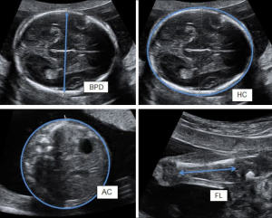
Fig- 2D Ultrasound
When are 2D ultrasounds normally done?
2D ultrasounds are commonly done for various medical and diagnostic purposes, including during pregnancy to monitor fetal development.
In the case of pregnancy, 2D ultrasounds are usually performed during the first trimester to confirm the pregnancy and estimate the gestational age, and then again during the second and third trimesters to monitor fetal growth and development, assess the placenta and amniotic fluid levels, and screen for certain fetal abnormalities.
Additionally, 2D ultrasounds may be used to diagnose or monitor various medical conditions that affect different organs in the body, such as the liver, kidneys, bladder, and reproductive organs.
The timing of 2D ultrasounds will depend on the specific medical condition being evaluated. In general, your healthcare provider will recommend a 2D ultrasound if they believe it is necessary to diagnose or monitor a condition. They will also provide instructions on when to schedule the ultrasound and whether any preparation is required before the procedure.
How is a 2D ultrasound done?
A 2D ultrasound is a non-invasive and painless procedure that uses high-frequency sound waves to create images of internal body structures. The steps of a 2D ultrasound may vary depending on the specific area of the body being examined, but the general process is as follows:-
a) Preparation:- You will typically be asked to change into a hospital gown and lie down on a table. The area of the body being examined will be exposed, and a gel will be applied to the skin. This gel helps to improve contact between the skin and the ultrasound probe and allows sound waves to pass through the skin more easily.
b) Ultrasound probe placement:- A handheld device called an ultrasound transducer is then placed on the skin over the area being examined. The transducer emits high-frequency sound waves, which pass through the skin and bounce back to the transducer as echoes. The echoes are then converted into an image by a computer.
c) Image capture:- The sonographer or ultrasound technician will move the transducer around the area being examined to capture images from different angles. They may ask you to change positions or take deep breaths to get a better view of the area being examined.
d) Image interpretation:- The images are then reviewed by a radiologist or other healthcare provider who interprets the results and provides a diagnosis.
e) Clean-up:- Once the procedure is complete, the gel will be wiped off, and you can return to your regular activities.
The entire procedure usually takes around 30 minutes, but it can take longer for more complex scans. You should not experience any pain during the procedure, although you may feel slight pressure from the transducer on your skin. If you have any concerns or questions about the procedure, you can speak with your healthcare provider or the sonographer performing the scan.
Is the weight 2D ultrasound standard?
No, weight measurement is not a standard part of a 2D ultrasound. A 2D ultrasound is primarily used to create images of internal body structures using high-frequency sound waves, and it is typically used in obstetrics to monitor fetal development during pregnancy, or for diagnosing and monitoring various medical conditions affecting different organs in the body.
While it is possible to estimate fetal weight using ultrasound, this is typically done using a different type of ultrasound called a biophysical profile, which combines 2D ultrasound with other measurements such as amniotic fluid levels, fetal breathing, fetal movements, and fetal heart rate. This type of ultrasound is typically done in the later stages of pregnancy to assess fetal well-being and to help determine if the baby needs to be delivered.
It is important to note that estimates of fetal weight using ultrasound can be imprecise, and may be affected by factors such as the position of the baby, the amount of amniotic fluid, and the size and shape of the mother’s pelvis. Your healthcare provider can provide more information on when and how fetal weight estimates are typically obtained during pregnancy.
How to calculate fetal weight through ultrasound?
Fetal weight can be estimated using ultrasound, but it is important to note that these estimates can be imprecise and can have a margin of error of up to 15% or more. Additionally, there are different formulas and methods that can be used to estimate fetal weight, and the accuracy can depend on a number of factors, including the baby’s position, the amount of amniotic fluid, and the skill of the sonographer performing the ultrasound.
One commonly used method to estimate fetal weight is the Hadlock formula, which uses measurements of the baby’s head circumference, abdominal circumference, and femur length to estimate fetal weight. The formula is as follows:
Estimated fetal weight (g) = 1.07 x (HC x AC x FL) + 1.57
Where
HC is the head circumference in millimeters.
AC is the abdominal circumference in millimeters.
FL is the femur length in millimeters.
The resulting estimated fetal weight is in grams.
It is important to note that fetal weight estimates are not always accurate, and they should not be used as the sole basis for making clinical decisions.

A student sonographer who needs assistance in learning